Raman and photoluminescence confocal microspectrometer.
Comprehensive physical & chemical microanalysis.
How it works
Raman spectroscopy is a non-destructive, non-contact and fast method for physical and chemical analysis which does not require any special sample preparation. The method allows to analyze solid, liquid, powdery samples, gases, solutions, suspensions, etc. No vacuum or other special conditions are required for measurements, spectra are usually recorded at atmospheric environment and room temperature.
The spectra obtained provide qualitative and quantitative information. Raman peaks positions indicate the chemical compound and its functional groups while the intensity indicates its concentration. An important information is also provided by shifts in peak positions with changes in temperature or pressure.
Together with infrared (IR) spectroscopy, Raman spectroscopy joins a group of techniques called vibrational spectroscopy. Both methods of spectroscopies study the same vibrational levels of molecules, but in different ways.
A Raman spectrometer integrated with an optical microscope (including a confocal one) is called “Raman microscope” or “Raman microspectrometer”; it has a number of important advantages. The excitation laser is focused into a small region near the microscope objective lens focus determined by the emission wavelength, the spectral Raman signal is emitted from the same region. It provides localized measurements with the possibility for the spectra being referred to various points on the sample surface.
Confocal Raman microspectroscopy with a small aperture provides high spatial resolution (sub-μm) and 3D mapping possibility. An optically transparent 3D sample can be studied layer by layer, and each thin layer of the sample can be explored separately with no need to make cross-sections and to destroy the sample.
The LS RamBo 620 microspectrometer is equipped with a high-precision motorized scanning stage and/ or motorized scanning mirrors for 2D/ 3D point-to-point mapping of spectra from a given area/ volume of the sample (Raman mapping).
In Raman spectroscopy it is possible to use low-power excitation lasers without a risk of the sample heating, the sample destruction, and the appearance of the thermal radiation parasitic lines. The spectra quality is unaffected by the atmospheric air, including the presence of water, so there is no need to dry or vacuumize the cuvette compartment. The spectra of aqueous solutions, including organic compounds, can be quantitatively analyzed without an uncontrollable absorption of the signal by an aqueous environment.
Applications
Raman confocal microspectrometers are widely used in science and technology. In pharmacy one may get tablet surface chemical images, i.e., to visualize the distribution of components, including depth profiling and optical sectioning. It is possible to perform rapid analysis of raw materials quality and authenticity, including working through transparent packaging without opening it, since polyethylene and other transparent polymers, as well as glass, have a very small Raman scattering cross-section. In the chemical laboratory, polymer synthesis and monomers conversion can be monitored in real time by spectral signal (spectrometers are equipped with the special optical fiber probes to control directly the reaction mixture).
In the semiconductor industry, it is possible to perform stress and strain mapping of silicon layers, to study the distribution of impurities and contaminants. Using an inverted optical microscope, it is possible to make 3D spectral maps of cells, including measurements in vivo, to study lipids, proteins, amino acids.
Due to the submicron resolution of modern confocal microscopes, Raman mapping is widely utilized in nanotechnology. It can be used to analyze the spectra of nanotubes, graphene layers, and other nanomaterials. In criminology, it is possible to perform a rapid non-destructive analysis of the sample chemical composition, e.g., to verify the authenticity of a written document. Raman spectrometers are also used in archaeology and art history to determine the exhibits authenticity and to identify the artifacts origin, the artistic pigments and the gemstones.
Design
The LS RamBo 620 spectrometer has the modular structure. A wide choice of excitation lasers and optical microscopes, automatic switching between excitation laser channels, automatic optical and mechanical parts adjustment to achieve maximum efficiency - all this makes the system flexible and solves a huge number of research tasks.
Stokes shifts from 100 cm-1 (optionally from 50 cm-1) can be measured. The monochromator measurement error limit by wavelength is 1 Å (grating 600 gr/mm).
The instrument is controlled by the LS RamBo software.
To work on the spectrometer, one may use various commercially available laser sources (with “free space” or fiber input type) and detectors (multi-channel or single-channel).
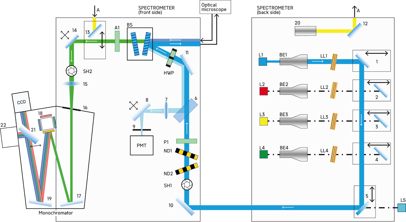
Simplified optical scheme of the LS RamBo 620 operation
L1, L2, L3, L4 - built-in lasers
L5 - external laser
BE1, BE2, BE3, BE4 - beam expanders
LL1, LL2, LL3, LL4 - laser line cleaning filters
ND1, ND2 - neutral density filter wheels
SH1, SH2 - shutters
BS - beam splitter unit (edge filters)
A1 - analyzer
P1 - polarizer
HWP - half-wave plates wheel
1, 2, 3, 4, 5, 13 - movable mirrors
10, 11, 12 – mirrors
6 (optional) - semi-transparent plate
7 (optional) - focusing optics
8 (optional) , 14 - motorized mirrors
9 (optional) - slit
16 - confocal aperture
17, 19 - concave mirrors
18 - diffraction grating unit (3 or 4 gratings on rotating turret)
20 - calibration lamp
21 (optional) - movable mirror
22 (optional) - second spectral detector
PMT (optional) - photomultiplier tube
The colors on the scheme
Laser radiation from source
Laser radiation scattered by the sample
Long-wavelength (Stokes) radiation component emitted by the sample. To realize Raman confocal measurements
Short-wavelength (anti-Stokes) radiation component emitted by the sample + laser radiation reflected from the sample. The laser line and the short-wavelength (anti-Stokes) radiation component intensities are quite different, so we assume the intensity of the reflected laser radiation is detected only, and the laser confocal measurement mode is realized
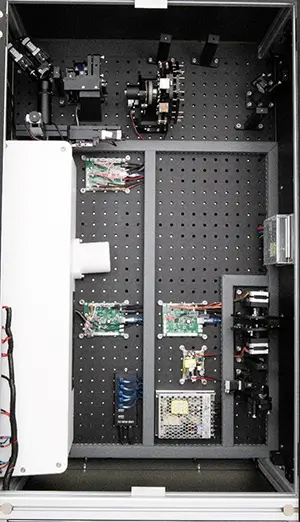
LS RamBo 620 front side with doors removed
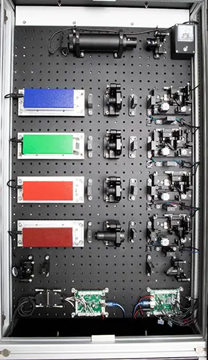
LS RamBo 620 back side with doors removed
Technical data
Diffraction gratings
up to 4 pcs. on the turret, to choose from: 150, 300, 600, 1200, 1800, 2400 gr/mm
Laser sources
Quantity
up to 5 (4 built-in + one external)
Power
up to 100 mW
Wavelength range
from 400 to 800 nm
Polarization
Linear
Beam profile
Gaussian TEM00, single longitudinal mode (SLM)
Monochromator entrance slit width
from 0 to 2000 µm, step – 1 µm
Edge/notch filters
8-position wheel, for filters Ø 12.5 mm and Ø 25 mm
Beam expander
optimized to fill the entrance pupil of the lens
Calibration lamp
two-element hollow cathode
Detectors
CCD detector PMT (optional) APD (optional)
LS RamBo 620 key features
Czerny-Turner spectrometer with 3 or 4 motorized diffraction gratings on a turret
Focal length 620 mm
Possibility to install up to 5 excitation lasers, internally or/ and externally
Possibility to install different detectors:
CCD detector to visualize the whole emission spectrum
APD or PMT (including photon counting mode) for weak signals
Motorized polarization optics in the excitation and emission channels
Automated switching between the excitation lines
Spatial resolution limited by the diffraction laws
Motorized confocal aperture for sensitivity and spatial resolution optimization
Beam expanders for complete filling of the pupil lens
 Laser radiation from source
 Laser radiation scattered by the sample
 Long-wavelength (Stokes) radiation component emitted by the sample. To realize Raman confocal measurements
 Short-wavelength (anti-Stokes) radiation component emitted by the sample + laser radiation reflected from the sample. The laser line and the short-wavelength (anti-Stokes) radiation component intensities are quite different, so we assume the intensity of the reflected laser radiation is detected only, and the laser confocal measurement mode is realized